Images
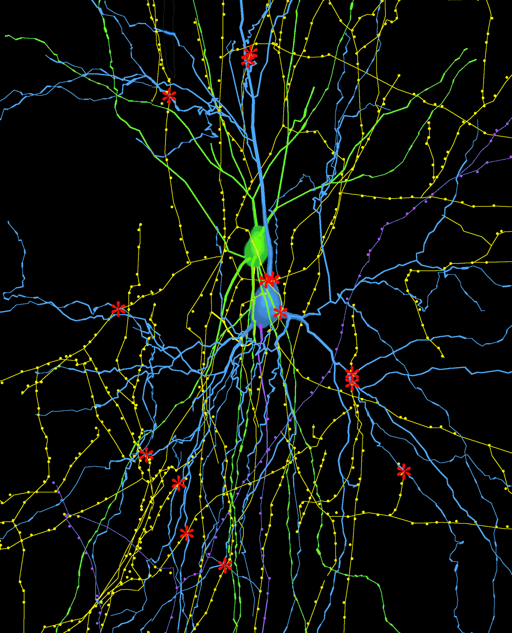
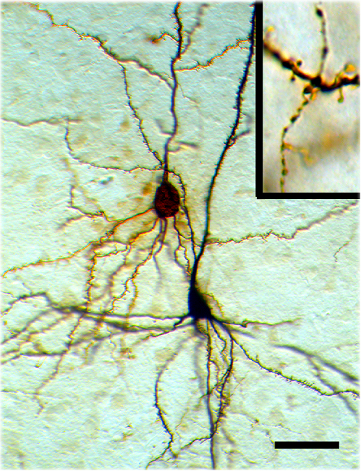
Synaptic connections between pyramidal neurons and GABAergic interneurons. The neurons were labeled with bicytin during patch clamp recording and reconstructed with Neurolucida system.
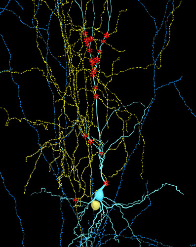
Lucifer Yellow labled pyramidal neuron and TH-ir fibers (blue)
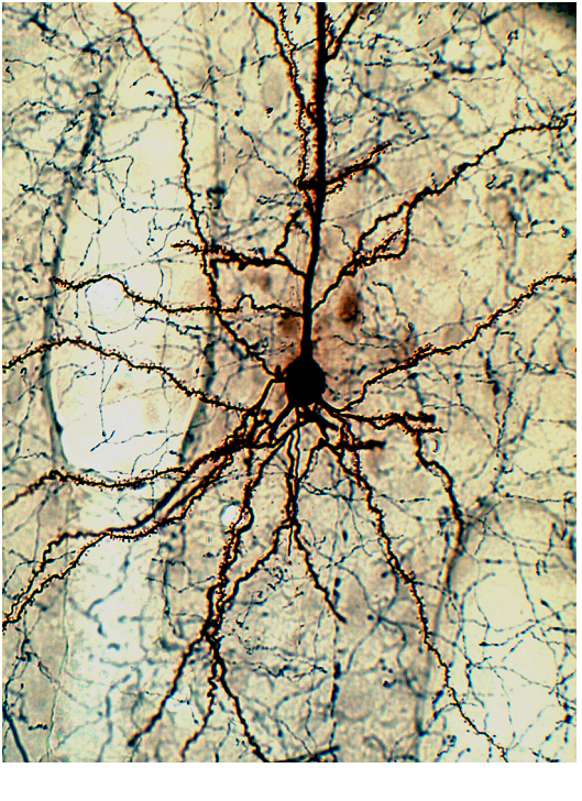
Biocytin-labeled layer 5 pyramidal neurons.
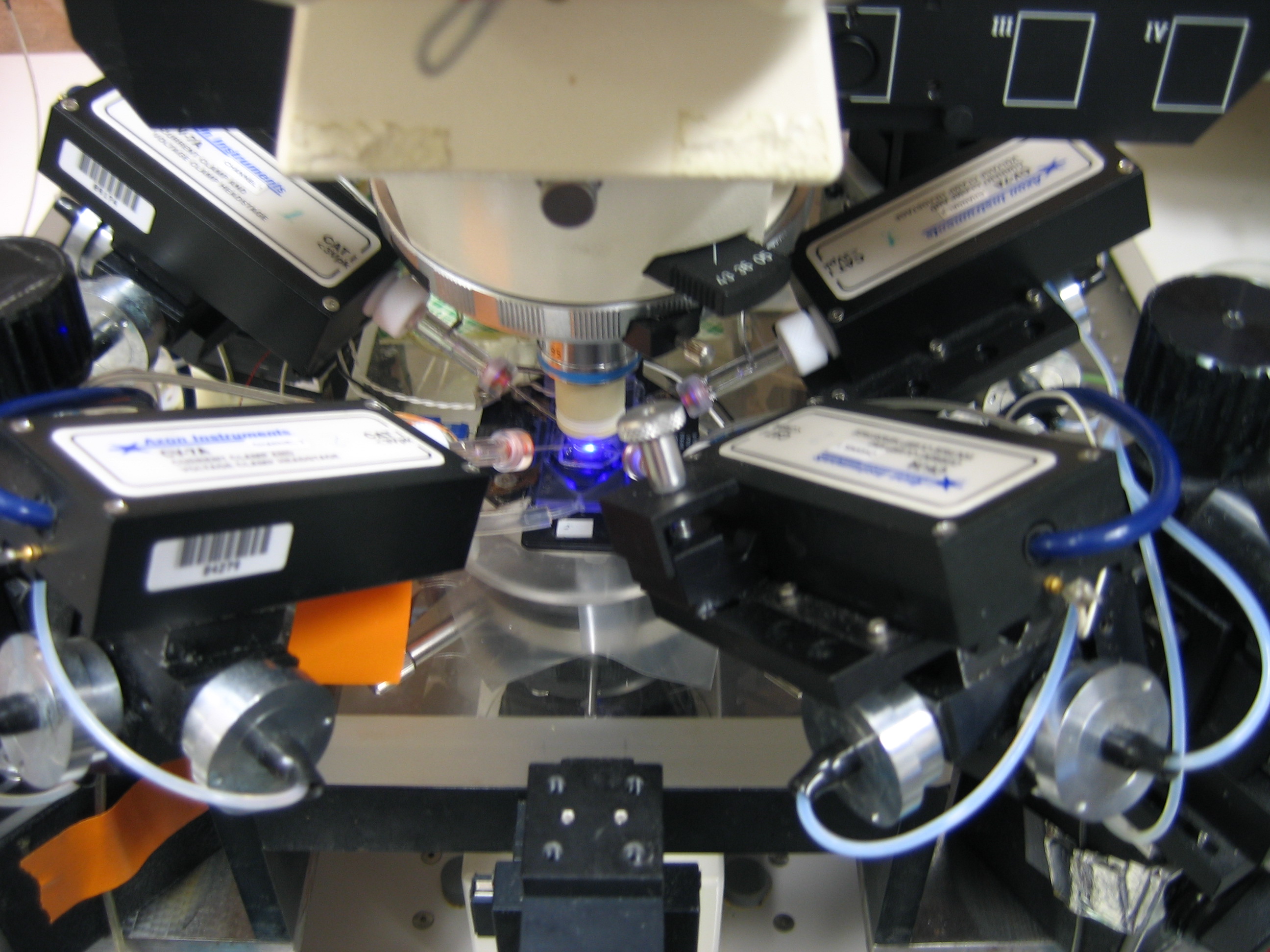
Setup for multiple whole-cell patch clamp recording.
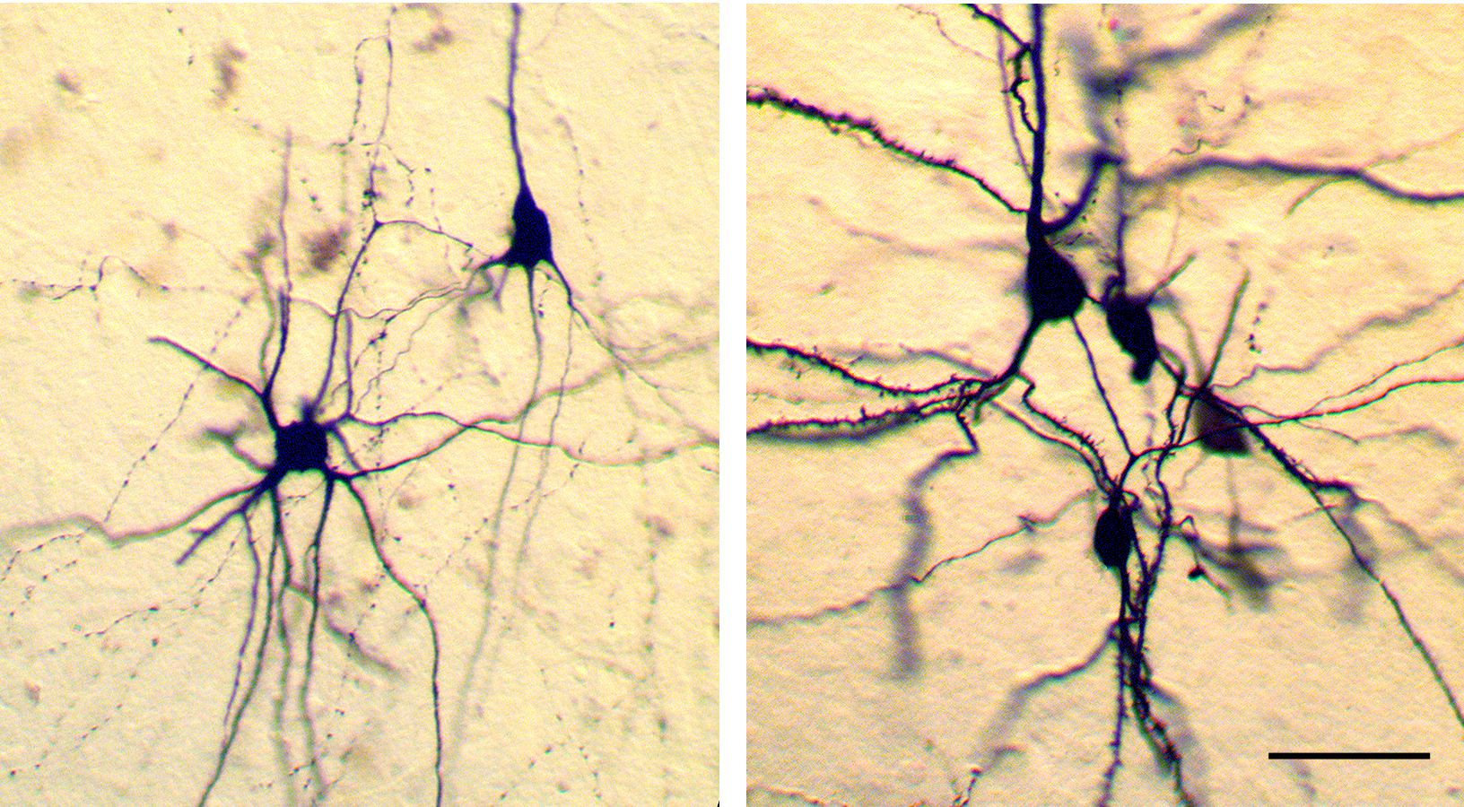
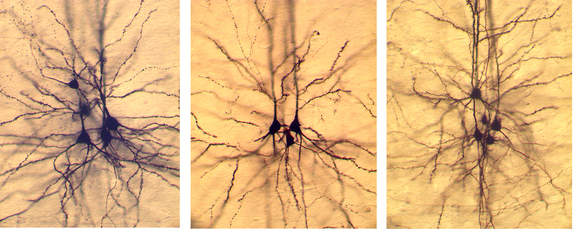
Biocytin-labeled interneurons and pyramidal neurons.
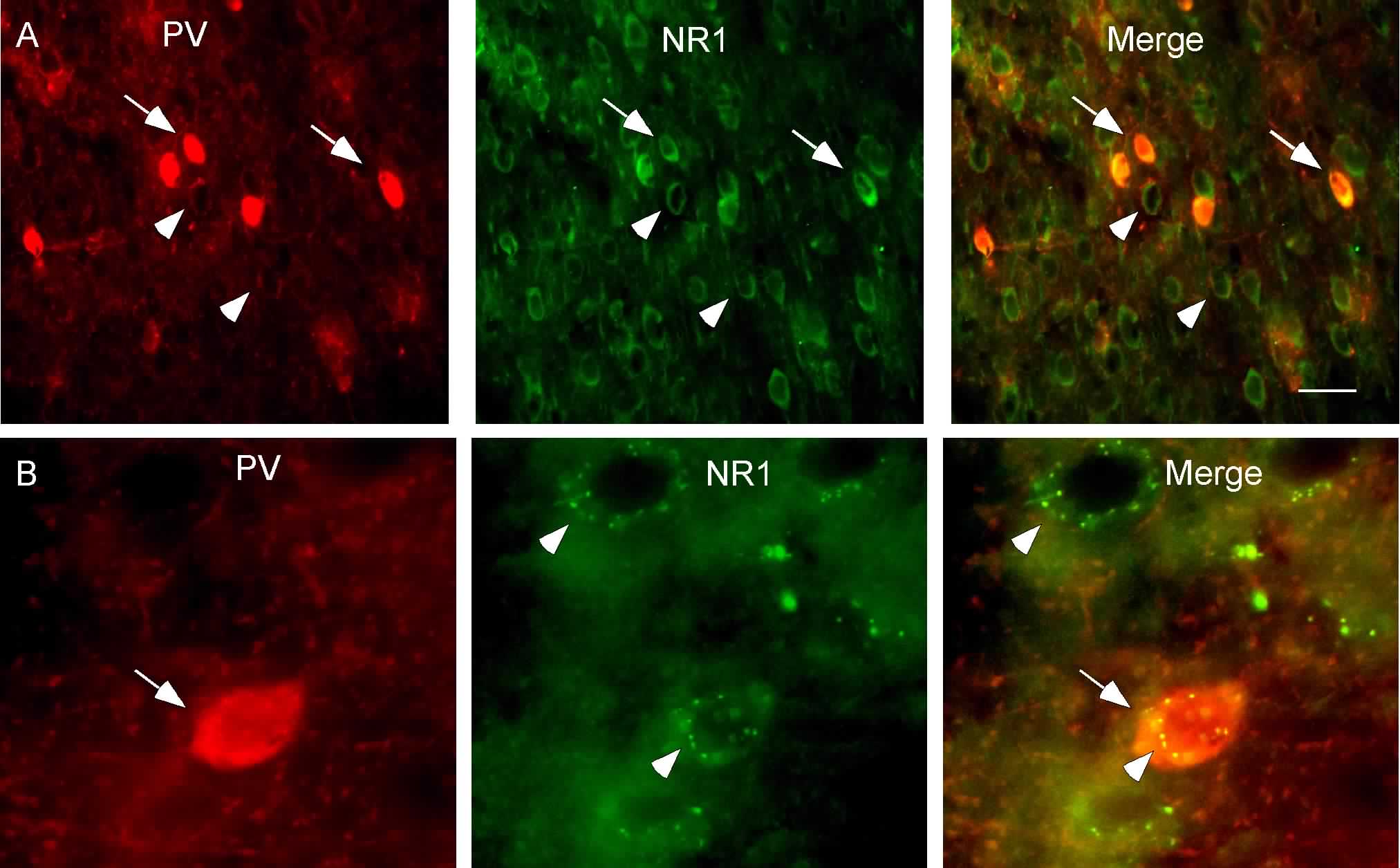
Double labeling of PV and NR1 subunits in the mPFC. PV, localized mainly in the fast-spiking interneurons in the neocortex, was labeled with Texas-Red (red) while NR1 was stained with FITC (green).
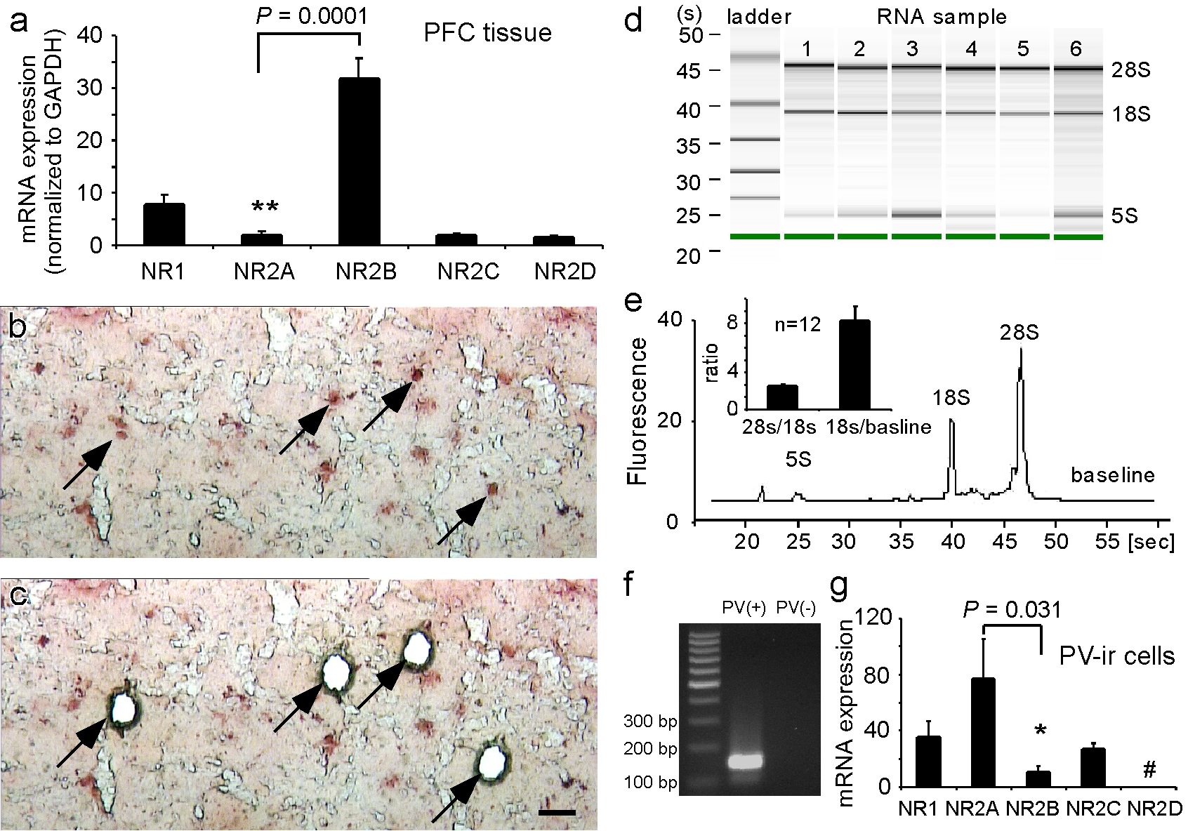
Relative mRNA expression of NMDAR subunits in adult rat PFC and parvalbumin-immunoreactive (PV-ir) interneurons. (a) mRNA expression of NMDAR subunits in the PFC tissue. NR2B mRNA expression is significantly higher than that of the NR2A subunit. (b, c) High-magnification photographs showing the PV-ir interneurons (arrows) labelled with rapid NovaRED immunostaining before (b) and after (c) the laser cut. Scale bar in (c)=50 um. (d, e) RNA integrity numbers (RIN) were measured with Agilent Bioanalyzer and the electropherogram of RNA extracted from laser microdissection (LMD)-picked PV-ir interneurons is shown in (d), where 5S covers the small rRNA fragments (5S and 5.8S rRNA and tRNA) and 18S and 28S cover the 18S peak and 28S peak (e). RIN=8.63 with 28S/18S=1.90 and 18S/baseline=7.23. ( f ) RT–PCR amplification of PV showing the expression of PV in LMD-captured PV-ir cells compared to that in PV-negative tissue. The left lane was the molecular weight marker with 100-bp intervals. (g) Relative mRNA expression of NMDAR subunits in PV-ir interneurons. The mRNA level of NR2A subunit was significantly higher than that of NR2B subunit.
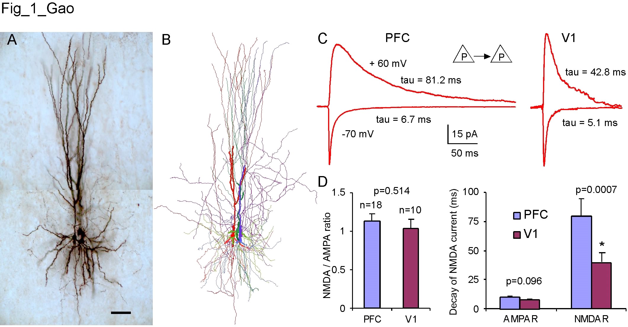
A and B, Biocytin-labeled layer 5 pyramidal neurons from multiple recordings and Neurolucida reconstruction, respectively; C and D, NMDA and AMPA receptor-mediaed currents recorded from the monosynaptic connections between pyramidal neurons in the adult rat prefrontal cortex.
Back to Top